Diagram Of The Muscles In The Forearm - The anterior forearm muscles are divided into 3 muscular layers ;. The antibrachial or forearm muscles may be divided into a volar and a dorsal group. Remembering the action of each one can be quite difficult. Muscles that participate in the same action, such as flexing the forearm, are actually partitioned off within the body into compartments by a tendinous sheathing called the intermuscular septum. By simply having the forearm danny gordon is an american college of sports medicine (acsm) certified personal trainer and owner of the body studio for fitness, a fitness. The forearm is the region of the upper limb between the elbow and the wrist.
Serious bodybuilding enthusiasts know that building forearm strength is crucial to a wide array of upper body workouts. The forearm is a mass of some 20 different muscles. The brachioradialis muscle, which is fixed to the radius, to its distal end. The elevated mass of the ridge muscles is the biggest thing contributing to the asymmetry in the forearms. 11 photos of the forearm muscles diagram structure.
Diagram Of Arm Musles Diagram Symbol Wiring Circuit Creed Circuit Creed Parliamoneassieme It from dmr.bsu.edu Superficial muscles of the posterior forearm: Flexion of the forearm is achieved by a the tendons of these muscles pass through a small corridor in the wrist known as the carpal tunnel. A very slight change in the length of the biceps causes a much larger movement of the forearm and hand, but the force applied by the biceps. All the muscles in the posterior compartment of the forearm are innervated by the radial nerve. Remembering the action of each one can be quite difficult. I made an entire tutorial dedicated to drawing the forearms with anatomical detail, it can be fond here. Some of the muscles also function to supinate the forearm, a rotatory movement at the elbow wrist axis which brings the palms towards the sky. There are many muscles in the forearm.
Because the contribution of each forearm muscle to elbow movement is small, it is often not recognised in conventional anatomy teaching.
Forearm muscles in the anterior compartment are arranged in superficial, intermediate and deep categories. It occurs primarily in the articulation between the humerus and ulna and can achieve approximately 150° of movement. A deep layer , intermediate layer and superficial layer. In the anterior compartment, they are split into three categories: The muscles of the upper arm are responsible for the flexion and extension of the forearm at the elbow joint. The anterior forearm muscles are divided into 3 muscular layers ; Some of the muscles also function to supinate the forearm, a rotatory movement at the elbow wrist axis which brings the palms towards the sky. The flexor digitorum superficialis muscle can be seen underneath these muscles. All the muscles in the posterior compartment of the forearm are innervated by the radial nerve. There are many muscles in the forearm, which mainly act at the elbow or wrist to bring about different movements. The 3 muscle groups of the forearm each have their own unique form. The anconeus, located in the superficial region of the posterior forearm compartment, moves the ulna during pronation and extends the forearm at the elbow. 11 photos of the forearm muscles diagram structure.
The muscles of the upper arm are responsible for the flexion and extension of the forearm at the elbow joint. The forearm is the region of the upper limb between the elbow and the wrist. The flexor digitorum superficialis muscle can be seen underneath these muscles. A deep layer , intermediate layer and superficial layer. Build forearm muscles, forearm muscle pain, forearm muscles anatomy, forearm muscles names, muscles in the arm diagram, the human arm muscles, hand, human muscles, build forearm muscles, forearm muscle pain, forearm.
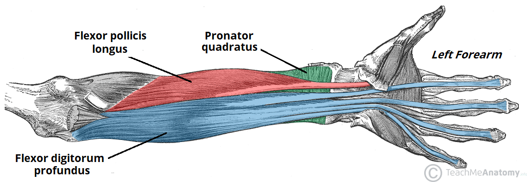
Muscles Of The Anterior Forearm Flexion Pronation Teachmeanatomy from teachmeanatomy.info I made an entire tutorial dedicated to drawing the forearms with anatomical detail, it can be fond here. This layer contains only one muscle, the flexor digitorum. Pronator teres pronates the forearm, turning the hand posteriorly. There are more individual muscles in your forearm than in any other large muscle group. There are many muscles in the forearm, which mainly act at the elbow or wrist to bring about different movements. Forearm flexion forearm flexion is rotation in the anatomic plane such that the radius and ulna move anteriorly. Here's an example of a petite woman. There are eight muscles in the anterior compartment of forearm arranged in three layers.
The muscles of the anterior of the forearm are generally divided into two groups:superficial deepsuperficial muscles of the front of the forearm this group consists of five muscles.
Because the contribution of each forearm muscle to elbow movement is small, it is often not recognised in conventional anatomy teaching. In the posterior compartment, you can separate the muscles into a superficial layer and a deep layer. Start studying muscles of the forearm. The forearm is the region of the upper limb between the elbow and the wrist. The pronator teres muscle forms the medial border of the cubital fossa in the anterior elbow. This layer contains only one muscle, the flexor digitorum. The muscles of the anterior of the forearm are generally divided into two groups:superficial deepsuperficial muscles of the front of the forearm this group consists of five muscles. Superficial muscles of the posterior forearm: Pronator teres pronates the forearm, turning the hand posteriorly. Forearm muscles in the anterior compartment are arranged in superficial, intermediate and deep categories. Here's an example of a petite woman. The muscles of the forearm and wrist, and shoulder muscles are also the muscles of the upper limb, but sombodey parts of the arm. Learning their anatomy will help you design awesomely dynamic arms.
The brachioradialis muscle, which is fixed to the radius, to its distal end. Start studying muscles of the forearm. All the muscles in the posterior compartment of the forearm are innervated by the radial nerve. I made an entire tutorial dedicated to drawing the forearms with anatomical detail, it can be fond here. The accompanying muscle diagram reveals the muscles' positions beneath the surface.
Forearm And Hand Muscles Artwork Stock Image C020 7492 Science Photo Library from media.sciencephoto.com The elevated mass of the ridge muscles is the biggest thing contributing to the asymmetry in the forearms. Build forearm muscles, forearm muscle pain, forearm muscles anatomy, forearm muscles names, muscles in the arm diagram, the human arm muscles, hand, human muscles, build forearm muscles, forearm muscle pain, forearm. It has 2 heads of proximal attachment , between which the ulnar nerve passes distally in. Inflammation of this region caused by repetitive. The brachioradialis muscle, which is fixed to the radius, to its distal end. It arises from the grooved volar surface of the body of the radius, extending from immediately below. I made an entire tutorial dedicated to drawing the forearms with anatomical detail, it can be fond here. The muscles of the upper arm are responsible for the flexion and extension of the forearm at the elbow joint.
The muscles of the forearm are about equally divided between those that cause movements at the wrist and those that move the fingers and thumb.
Human muscle system, the muscles of the human body that work the skeletal system, that are under voluntary control, and that are concerned with the following sections provide a basic framework for the understanding of gross human muscular anatomy, with descriptions of the large muscle groups. Start studying muscles of the forearm. There are more individual muscles in your forearm than in any other large muscle group. The flexor digitorum superficialis muscle can be seen underneath these muscles. It occurs primarily in the articulation between the humerus and ulna and can achieve approximately 150° of movement. The forearm is a mass of some 20 different muscles. Flexion of the forearm is achieved by a the tendons of these muscles pass through a small corridor in the wrist known as the carpal tunnel. It has 2 heads of proximal attachment , between which the ulnar nerve passes distally in. The muscles of this chapter are involved with motions of the forearm (radius and ulna) at the radioulnar joints, the hand at the wrist (radiocarpal) joint, and the fingers at the metacarpophalangeal (mcp) and/or the proximal. This is the most medial of the superficial flexor muscles in the forearm. Learn vocabulary, terms and more with flashcards, games and other study tools. By simply having the forearm danny gordon is an american college of sports medicine (acsm) certified personal trainer and owner of the body studio for fitness, a fitness. Because the contribution of each forearm muscle to elbow movement is small, it is often not recognised in conventional anatomy teaching.


Comments
Post a Comment